
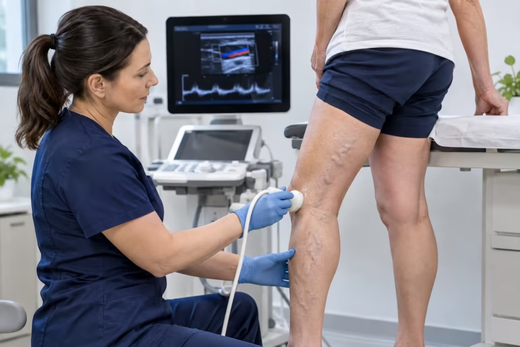
A venous duplex ultrasound is a non-invasive scan that examines the veins in your legs and measures blood flow. For varicose veins, it helps identify faulty valves, backward blood flow (venous reflux) and affected veins. During the examination, a sonographer moves an ultrasound probe along your leg while you stand or change position. The scan requires no needles or radiation.
The results show more than the veins visible beneath your skin. They help your clinician assess the circulation within the leg and determine whether further treatment needs to be considered.
What Is a Venous Duplex Ultrasound?
A venous duplex ultrasound combines conventional ultrasound images with Doppler measurements of blood flow.
The images show the structure and course of veins, while Doppler technology measures the direction and characteristics of blood movement.
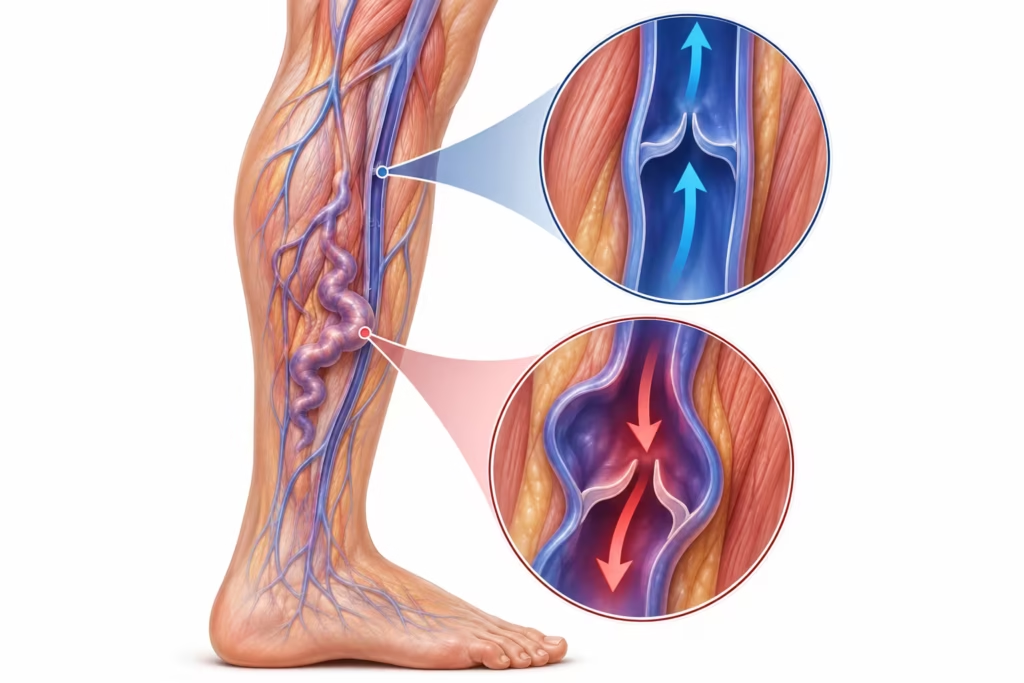
Healthy leg veins contain one-way valves that help blood return towards the heart. If these valves stop closing effectively, blood can travel backwards and contribute to increased venous pressure.
This is known as venous reflux.
Duplex ultrasound is used to investigate the location and extent of reflux, particularly in patients experiencing symptomatic or recurrent varicose veins.
Venous Duplex Ultrasound: Key Facts
| Question | What patients should know |
|---|---|
| What does the scan examine? | Vein anatomy, blood flow, valve function and reflux |
| Is it invasive? | No needles or incisions are needed |
| Does it use radiation? | No |
| Will I need to stand? | Often, particularly for reflux assessment |
| Is it painful? | Usually not, although pressure over tender areas may cause discomfort |
| Do I need to fast? | Usually not for a standard leg vein scan |
| How long does it take? | Depends on the scan requested and complexity |
| What happens to the results? | Findings are recorded and reported to the referring or treating clinician |
The NICE guideline on varicose veins recommends duplex ultrasound to confirm varicose vein disease, assess truncal reflux and plan treatment where appropriate.
What Happens During a Venous Duplex Ultrasound for Varicose Veins?
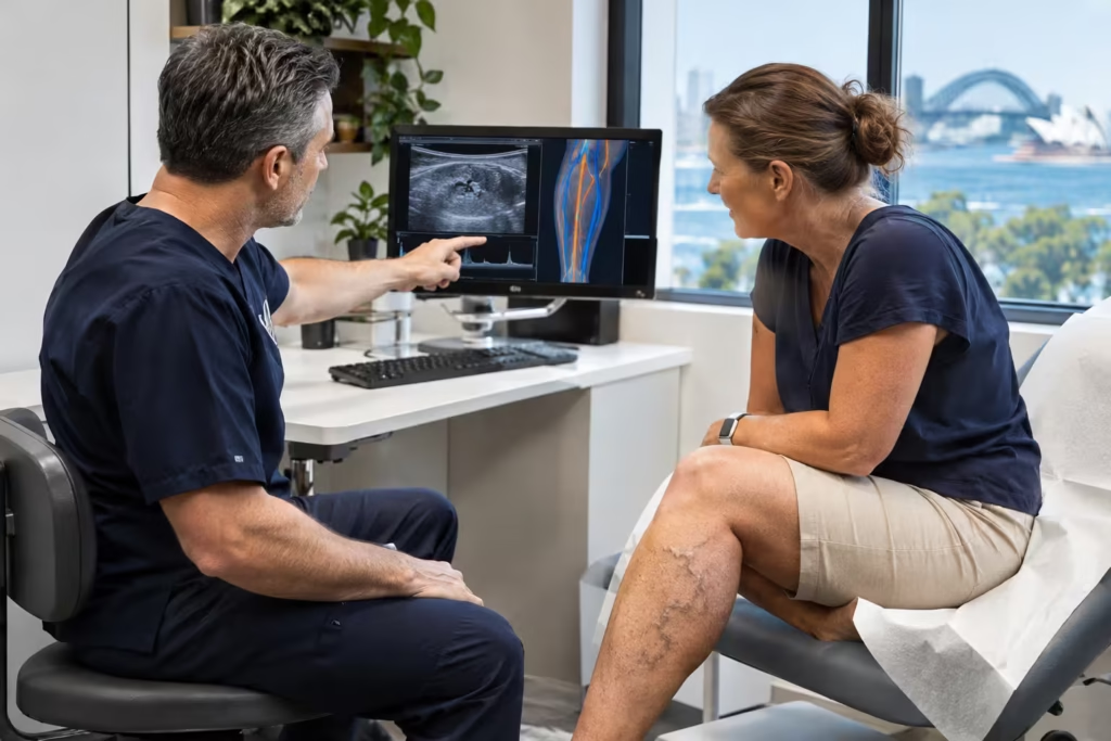
The examination follows the course of selected veins while the sonographer checks how blood flows through them.
The precise protocol depends on the clinical question, but a varicose vein reflux study typically includes the following steps.
1. Reviewing Your Symptoms and Medical History
Before scanning, the sonographer may ask about:
- Leg aching, heaviness or swelling
- Visible or painful varicose veins
- Previous vein treatment
- A history of blood clots
- Skin changes or leg ulcers
- Previous ultrasound examinations
This information helps guide which veins require detailed examination.
Bring relevant previous imaging reports if available.
2. Positioning Your Leg
You may be asked to stand during a venous reflux ultrasound.
Standing allows gravity to influence blood flow, making it easier to examine valves for abnormal backward flow.
The sonographer may also ask you to change position or shift your weight.
If standing is uncomfortable or you experience dizziness, tell the sonographer so another suitable position can be considered.
3. Applying Gel and the Ultrasound Probe
Water-based ultrasound gel is applied to the skin.
The sonographer then moves a handheld probe along relevant areas of the leg, including the calf, knee, thigh or groin.
The probe creates images of the veins beneath the skin.
Some pressure may be necessary to obtain clear images. Tell the sonographer if an area is particularly painful.
4. Testing Blood Flow and Vein Valves
During the examination, the sonographer may gently compress and release parts of the leg or ask you to perform a short breathing manoeuvre.
These actions temporarily change blood flow.
The Doppler component records how blood responds, including whether it travels backwards through valves that are not functioning properly.
The duration and location of backward flow help the clinician assess the significance of the findings.
5. Recording Vein Measurements
When the scan is being used for treatment planning, selected veins may be measured and mapped.
The sonographer records relevant anatomy, vein diameter and areas of reflux.
This creates a detailed record for the interpreting clinician rather than relying solely on the appearance of surface veins.
A report is then prepared for the referring or treating practitioner.
How Long Does a Varicose Vein Ultrasound Take?
The time needed for a venous duplex ultrasound varies with the examination requested.
Scanning both legs, mapping multiple veins or investigating previously treated veins can require additional measurements.
Confirm the expected appointment duration directly with the imaging provider so you can plan accordingly.
How Should You Prepare?
Most lower-limb venous duplex examinations require minimal preparation.
Before attending:
- Wear loose clothing that allows access to your legs.
- Bring your imaging request and relevant previous reports.
- Tell the provider about previous blood clots or vein procedures.
- Ask whether compression stockings should be removed beforehand.
- Mention any difficulty standing for extended periods.
Fasting is generally unnecessary for a standard lower-limb venous duplex ultrasound unless the provider instructs otherwise.
Does a Venous Duplex Ultrasound Hurt?
The scan itself usually causes little discomfort.
There are no needles, injections or incisions. The sonographer uses a probe on the skin, occasionally applying pressure to assess a vein or obtain clearer images.
If your leg is already tender or swollen, this pressure may feel uncomfortable.
Let the sonographer know if you experience pain, dizziness or difficulty maintaining the required position.
Venous Reflux Ultrasound vs DVT Ultrasound
Both examinations use duplex ultrasound technology, but they answer different clinical questions.
| Examination | Main purpose |
|---|---|
| Venous reflux ultrasound | Investigates chronic valve dysfunction and abnormal backward blood flow |
| DVT ultrasound | Investigates possible blood clots in the deep veins |
| Vein mapping ultrasound | Records relevant vein anatomy for assessment or treatment planning |
A venous reflux study and a DVT ultrasound are not automatically interchangeable.
A reflux study primarily investigates chronic venous disease. Suspected deep vein thrombosis requires an appropriate blood-clot assessment protocol.
If you develop sudden leg swelling or pain, do not wait for a routine varicose vein scan.
What Do Your Venous Duplex Ultrasound Results Mean?
The report describes the veins examined, their structure and the direction of blood flow.
Several findings may be relevant to varicose vein management.
Venous Reflux
Venous reflux occurs when blood flows backwards through a vein because its valves are not working effectively.
The report may identify which veins are affected and how long the backward flow continues.
For certain superficial veins, reflux lasting more than 0.5 seconds is a commonly used threshold. Different vein types and clinical circumstances have different interpretation criteria.
Finding reflux does not automatically mean a procedure is required.
Enlarged or Abnormal Veins
The examination may record the diameter, location and course of abnormal veins.
These measurements can help identify the relationship between visible varicose veins and the underlying venous circulation.
No Significant Reflux
A scan that does not identify significant reflux may lead the clinician to consider other explanations for leg discomfort or swelling.
Symptoms such as heaviness, pain and swelling are not exclusive to varicose vein disease.
The next step is determined by the full clinical assessment, not a single ultrasound measurement.
How Reflux Mapping Helps Guide Treatment
One important contribution of duplex ultrasound is identifying where abnormal blood flow originates, rather than simply recording which veins appear enlarged.
For example, reflux involving a major superficial vein may require a different management discussion from a smaller, isolated abnormal vein.
The clinician also examines whether the ultrasound findings correspond with the patient’s symptoms and skin changes.
The assessment may lead to a discussion about conservative management, sclerotherapy, endovenous treatment or surgery, according to the individual case.
Clinical guidelines support duplex ultrasound as part of treatment planning for suspected primary or recurrent varicose veins.
Is a Venous Duplex Ultrasound Covered by Medicare in Australia?
A venous duplex ultrasound may qualify for a Medicare rebate when the examination meets the requirements of an applicable Medicare Benefits Schedule (MBS) item.
Eligibility depends on the clinical indication, examination performed, valid imaging request and relevant billing requirements.
Relevant Medicare Items
| MBS item | Description |
|---|---|
| 55246 | Specified unilateral duplex scanning of lower-limb veins for chronic venous disease |
| 55296 | Specified unilateral duplex scanning and vein marking before varicose vein surgery, including associated skin marking |
Item 55296 is specifically related to preoperative vein marking. It does not apply automatically to every vein-mapping examination.
The imaging provider determines which item, if any, applies to the service provided.
Official references:
Do You Need a Referral?
A request for diagnostic imaging and a referral for a specialist consultation are different documents.
Medicare-funded diagnostic imaging generally requires a valid request from an appropriate healthcare practitioner, subject to the relevant MBS rules.
If you have been advised to undergo a venous duplex scan, confirm that the request specifies the examination needed.
Will There Be an Out-of-Pocket Cost?
A Medicare rebate does not necessarily cover the full cost.
Before booking, ask:
- Is my examination eligible for Medicare?
- Will the scan be bulk billed?
- What is the expected out-of-pocket fee?
- Does the request cover one leg or both?
- Is a reflux study or detailed vein mapping included?
Medicare information should be checked against the current MBS at the time of the examination.
When Should Varicose Veins Be Assessed?
A medical assessment is advisable when varicose veins cause persistent symptoms or are associated with changes in the skin.
Reasons to seek advice include:
- Recurrent leg aching or heaviness
- Swelling, especially after prolonged standing
- Painful or prominent varicose veins
- Skin discolouration or eczema-like changes near the ankles
- Symptoms returning after previous vein treatment
- A lower-leg wound that is not healing
These symptoms help determine whether further venous investigation is warranted.
Warning Signs Requiring Urgent Medical Care
Sudden unexplained swelling in one leg, especially with pain, warmth or redness, requires prompt assessment for possible DVT.
Call 000 in Australia for sudden chest pain, severe breathlessness, collapse or symptoms suggesting pulmonary embolism.
Bleeding from a varicose vein also requires immediate attention. Apply firm direct pressure, elevate the leg if possible and seek urgent medical help.
Varicose Vein Assessment at HealthiClinic in Greenwich, Sydney
HealthiClinic provides phlebology consultations for patients with varicose veins, spider veins, chronic venous insufficiency and other venous concerns.
A consultation can help determine whether symptoms suggest underlying venous reflux and whether duplex ultrasound is appropriate.
Clinical Expertise: Dr Joseph Gracé
Dr Joseph Gracé is a phlebologist at HealthiClinic in Greenwich, Sydney.
His published clinical areas include:
- Varicose and spider vein assessment
- Chronic venous insufficiency
- Venous ultrasound evaluation
- Sclerotherapy
- Minimally invasive endovenous procedures
Venous duplex ultrasound can identify the source and extent of reflux that may not be apparent from visible varicose veins.
This information helps the treating clinician determine whether the ultrasound findings explain the patient’s symptoms and which management options warrant discussion.
In Dr Gracé’s area of practice, these findings are relevant to deciding whether conservative measures, additional investigations or venous treatment should be considered.
An important clinical distinction: The location of visible veins does not necessarily reveal the origin of abnormal blood flow. Mapping the affected venous system helps connect the external findings with the underlying circulation.
This is a general evidence-based clinical explanation, not a quotation or personal case observation attributed to Dr Gracé.
His published professional profile describes his work in venous assessment and minimally invasive treatment.
Read Dr Joseph Gracé’s HealthiClinic profile.
Arranging an Appointment
Patients can contact HealthiClinic to confirm:
- Availability of phlebology consultations
- Referral requirements
- Whether ultrasound is performed onsite or organised externally
- Any required imaging requests
- Consultation and examination costs
Learn about HealthiClinic’s phlebology services.
Clinical attribution: Dr Gracé’s professional experience is supported by his published practitioner profile. His review of this article has not yet been confirmed.
Your ultrasound report is reviewed alongside your symptoms, medical history and clinical examination.
If reflux or another abnormality is identified, your clinician can explain whether monitoring, conservative care or further treatment is appropriate.
The report may also indicate that another cause of your symptoms needs investigation.
HealthiClinic provides phlebology consultations in Greenwich, Sydney, for people experiencing symptomatic varicose veins and other venous concerns.
For information about assessment and appointments:
- HealthiClinic Phlebology Services
- Dr Joseph Gracé — Phlebologist
- Vascular Surgery Services
- Contact HealthiClinic
Medical References and Clinical Sources
- National Institute for Health and Care Excellence (NICE). Varicose Veins: Diagnosis and Management (CG168). Guidance on venous assessment, duplex ultrasound and treatment planning.
- Society for Vascular Surgery, American Venous Forum and American Vein and Lymphatic Society. Clinical Practice Guidelines for Varicose Veins. Evidence-based guidance on venous reflux investigation and management.
- Australian Government — Medicare Benefits Schedule. Item 55246. Unilateral lower-limb duplex ultrasound for chronic venous disease.
- Australian Government — Medicare Benefits Schedule. Item 55296. Unilateral preoperative duplex scanning and vein marking.
- HealthiClinic. Dr Joseph Gracé — Practitioner Profile. Published professional scope and expertise.
- HealthiClinic. Phlebology Services. Information about venous assessment and treatment services.
Publisher: HealthiClinic
Prepared: 10 November 2026
Clinical reviewer: Pending confirmation
Medical disclaimer: This article provides general educational information and does not replace personal medical advice, diagnosis or treatment. Seek urgent medical care for suspected blood clots, significant bleeding or other vascular emergencies.
Do I have to stand during a venous duplex ultrasound?
Standing is commonly used for venous reflux assessment because gravity helps reveal backward blood flow. If you cannot stand comfortably, the sonographer may use another suitable position.
Can a venous duplex ultrasound detect blood clots?
Yes. Duplex ultrasound is used to investigate suspected DVT, but a DVT study follows a different examination protocol from routine varicose vein reflux mapping. Sudden leg symptoms need prompt medical assessment.
What happens if my ultrasound shows venous reflux?
Your treating clinician compares the reflux findings with your symptoms, examination and medical history. Management may include monitoring, conservative treatment or a venous procedure, depending on your circumstances.
Do I need to stop wearing compression stockings before the scan?
Ask the imaging provider when booking. You will generally need the relevant areas of the leg accessible during scanning, but the provider should advise when to remove compression garments.
Does Medicare cover a venous duplex ultrasound?
Some examinations qualify for Medicare rebates when the relevant MBS criteria are met. Eligibility depends on the scan performed and applicable billing requirements. Confirm the rebate and any gap fee with the provider.
What Happens After Your Venous Duplex Ultrasound?
If reflux or another abnormality is identified, your clinician can explain whether monitoring, conservative care or further treatment is appropriate.